CT scans combine multiple X-ray images into “slices” — detailed, cross-sectional images of bones, blood vessels and soft tissues. During a CT scan, you will lie on a table while a CT scanner moves around you, taking pictures.
CT scans have become the go-to exam for many physicians, as they can provide a wealth of information in a relatively short time. Since so much information can be obtained from a single image, CT scans can limit additional imaging and radiation exposure. And, as one of the quickest imaging methods, CT scans are a favorite tool among emergency physicians.
In the Stroke and Trauma programs, CT scans are key components of treatment:
- Head CT without contrast: This is the main mode of imaging during an acute stroke. It allows the physician to identify early signs of a stroke and exclude other conditions that could be mimicking a stroke, such as a tumor or bleeding within the skull.
- CT perfusion: This CT provides information about blood flow to the brain, including which areas are receiving enough blood. CT perfusion helps determine whether or not you are a candidate for having a blood clot removed from your head.
Some CT scans are used for screening procedures. For example, if you have a high risk of lung cancer, a low-dose CT can screen for signs of cancer. Or, if you have a risk of coronary artery disease, your physician may order a CT calcium score exam to check for a buildup of plaque in your coronary arteries.
We are also able to use CT technology when we perform several types of procedures:
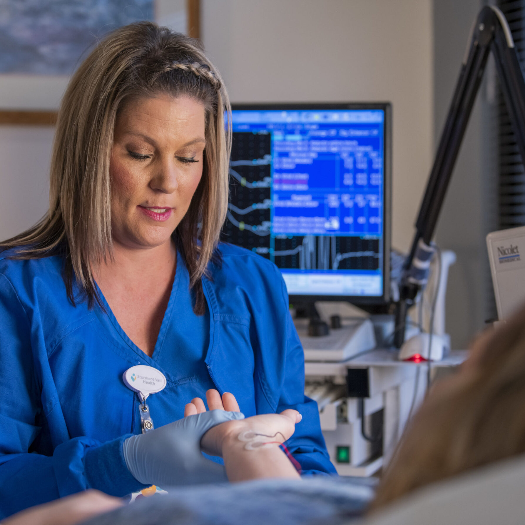
- Biopsy: A biopsy is a procedure where we remove a sample of tissue or cells from your body in order for it to be examined in a laboratory. In a CT-guided biopsy, we use imaging technology to guide a needle to an area of your body that needs a biopsy, but isn’t easily accessible through the skin (e.g., liver, lung).
- Chest tube placement: A chest tube is used to drain excess fluid, blood or air from around your heart, lungs or esophagus. We can use CT imaging to guide the tube to the correct location, meaning we can place the tube without performing open surgery.
- Radiofrequency ablation: Using CT imaging as a guide, we deliver heat and electric energy to cancerous or precancerous cells. This kills the cells, with only minimal impact to surrounding tissue.